The human brain, the three-pound conductor of our thoughts, emotions, and actions, has long been shrouded in mystery. Scientists have tirelessly endeavored to unravel its intricate workings, but its sheer complexity has presented a formidable challenge.
However, recent advancements in artificial intelligence (AI) are offering a powerful new lens through which we can observe this remarkable organ.
In a groundbreaking collaboration between Google researchers and Harvard neuroscientists, AI has been instrumental in generating a series of incredibly detailed images of the human brain. These images provide unprecedented views into the brain’s structure, offering a glimpse into the labyrinthine network of neurons that underlies our very existence.
A million gigabytes for a millionth of a brain
Imagine peering into a universe contained within a universe. This analogy aptly describes the challenge of studying the human brain. Its structure is mind-bogglingly intricate, composed of billions of neurons interconnected by trillions of synapses. To gain a deeper understanding, researchers require incredibly detailed information.
The research team used advanced AI tools to analyze a tiny sample of human brain tissue, specifically a section of the cortex from the anterior temporal lobe. This sample, though minuscule – representing only about one-millionth of the entire brain – contained a staggering amount of information. Astonishingly, processing this data required a mind-numbing 1.4 petabytes of storage, which is equivalent to over a million gigabytes.
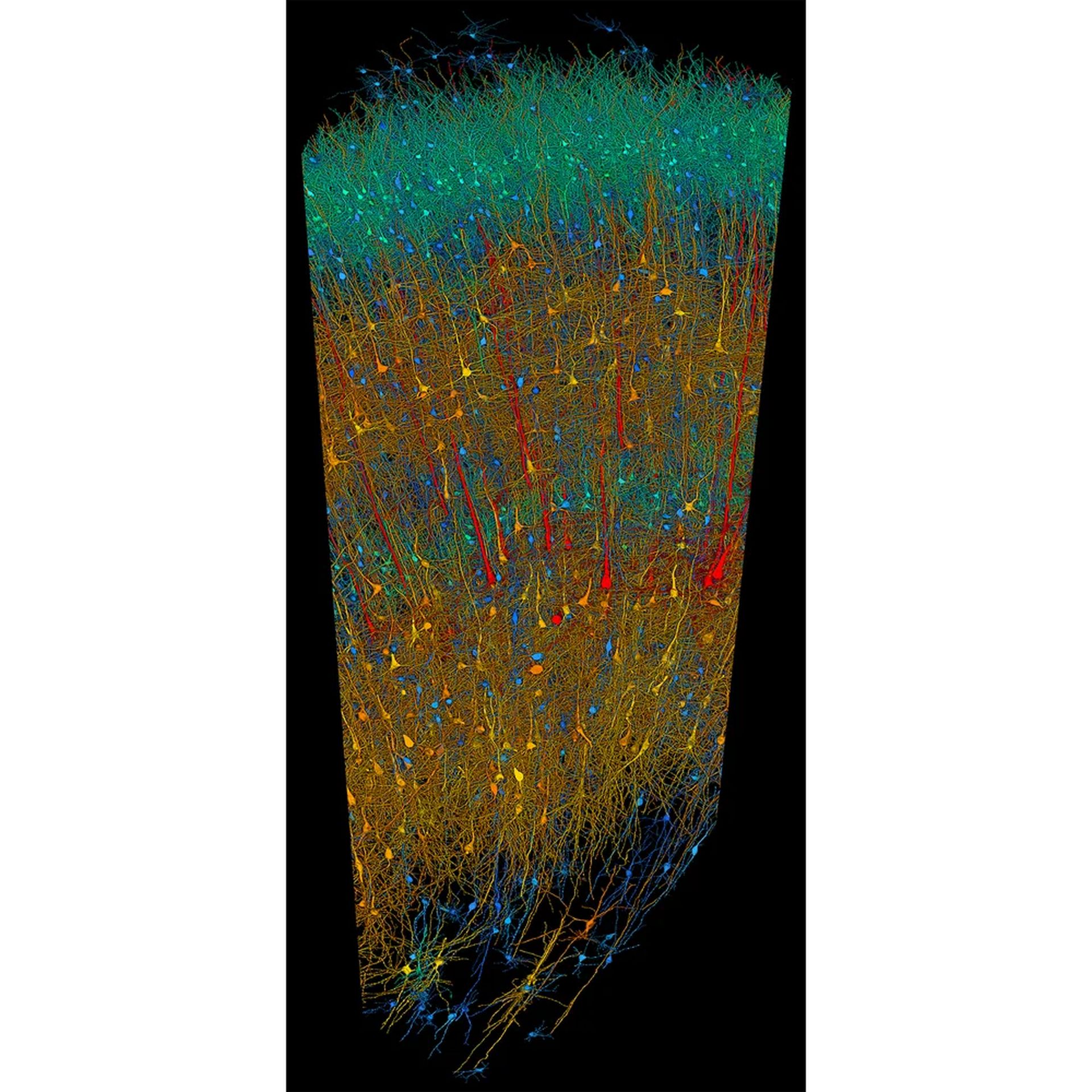
The AI processed this data to construct a high-resolution, three-dimensional model of the brain tissue. This model allows scientists to virtually navigate the intricate folds and layers of the brain, examining individual neurons and their connections in unparalleled detail.
Our cortex is a six-layered marvel
The outermost layer of the brain, the cortex, is responsible for our most complex cognitive functions, including language, memory, and sensory perception. This region is further divided into six distinct layers, each with a specialized role.
One of the most remarkable images generated by Google’s AI offers a zoomed-out view of all the neurons within the sample tissue. By coloring the neurons based on their size and type, the image reveals the distinct layering of the cortex. This visualization allows scientists to study how different types of neurons are distributed throughout the cortex and how they might contribute to specific functions.
Further analysis of the individual layers can provide even more granular insights. By zooming in, researchers can examine the intricate connections between neurons within each layer. These connections, known as synapses, are the fundamental units of communication in the brain. Understanding the organization of these connections is crucial for deciphering how information flows through the brain and how different brain regions interact with each other.

Mapping the myriad of neurons
The human brain is estimated to contain roughly 86 billion neurons, each with a unique role to play. These neurons come in a variety of shapes and sizes, and their specific characteristics influence how they transmit information.
Another image generated by Google’s AI provides a detailed census of the different types of neurons present within the sample tissue. By classifying the neurons based on their morphology, the researchers can begin to understand the relative abundance of different neuronal types in this specific brain region. This information can be compared to data from other brain regions to identify potential variations in neuronal composition across different functional areas.
Furthermore, AI can be used to analyze the spatial distribution of these different neuronal types. Are certain types of neurons clustered together, or are they more evenly dispersed throughout the tissue? Understanding these spatial patterns can shed light on how different neuronal populations interact with each other to form functional circuits within the brain.
Zooming into the dendrites and axons
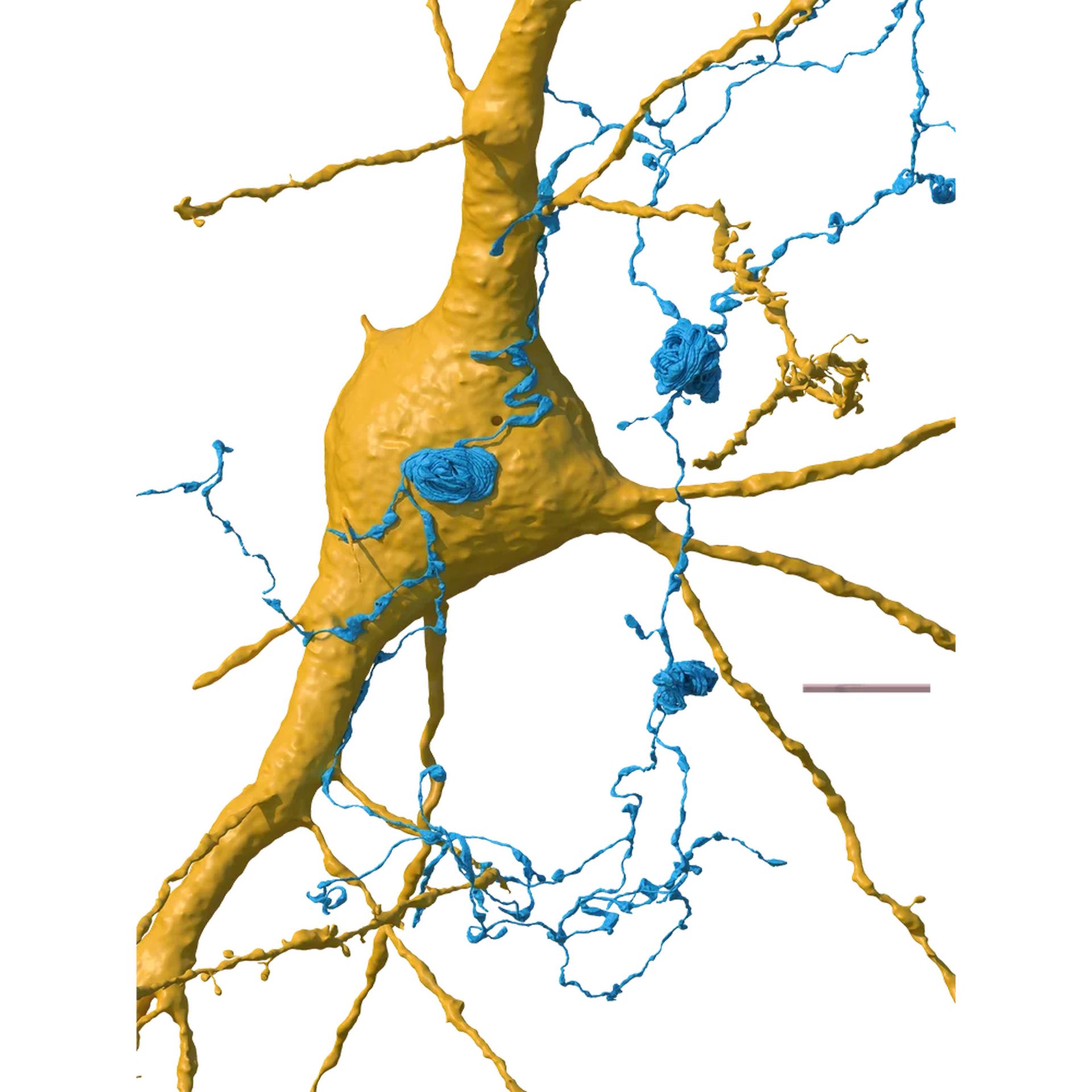
The magic of the brain lies in its ability to transmit information across vast networks of neurons. This communication occurs through specialized structures called dendrites and axons. Dendrites are like tiny antennae that receive signals from other neurons, while axons act as long, slender cables that transmit signals away from the cell body.
One of the most captivating images generated by Google’s AI provides a close-up view of the intricate dance of dendrites and axons within the sample tissue. This image allows researchers to visualize the density of these structures and how they connect with each other. The complex branching patterns of dendrites and the long, winding paths of axons reveal the intricate web of communication that takes place within the brain.
By analyzing the connectivity patterns, scientists can begin to map the functional circuits that underlie specific brain functions. They can identify groups of neurons that are likely to be involved in processing similar types of information and trace the pathways through which information flows from one brain region to another.

A new window into the human mind
The images generated by Google’s AI are a huge step for our ability to study the human brain. The detailed visualizations offer a window into the brain’s intricate structure, providing a wealth of information about the organization of neurons, their connections, and the cellular diversity within specific brain regions.
This newfound ability to explore the brain at such a granular level has the potential to revolutionize our understanding of neurological and psychiatric disorders. We already know AI’s potential for curing Alzheimer’s and by comparing brain tissue samples from healthy individuals to those with various conditions, researchers can identify potential abnormalities in neuronal structure or connectivity.
Furthermore, AI-generated images can be used to study the effects of aging, learning, and experience on the brain. By examining how the brain’s structure changes over time or in response to different stimuli, researchers can gain valuable insights into the mechanisms that underlie these processes.
The potential applications of this technology extend beyond the realm of basic research. The detailed models of brain tissue generated by AI could be used to develop more realistic simulations of brain function. These simulations could be used to test the effects of potential drugs or therapies before they are administered to human patients.
The vast majority of the brain remains uncharted territory, and many fundamental questions about its function continue to baffle scientists. However, these initial images offer a powerful glimpse into the brain’s hidden depths, and they pave the way for a future where we can finally begin to unravel the mysteries of this most complex organ.
Featured image credit: vecstock/Freepik


